
Proximal Tibia/Fibula Fracture
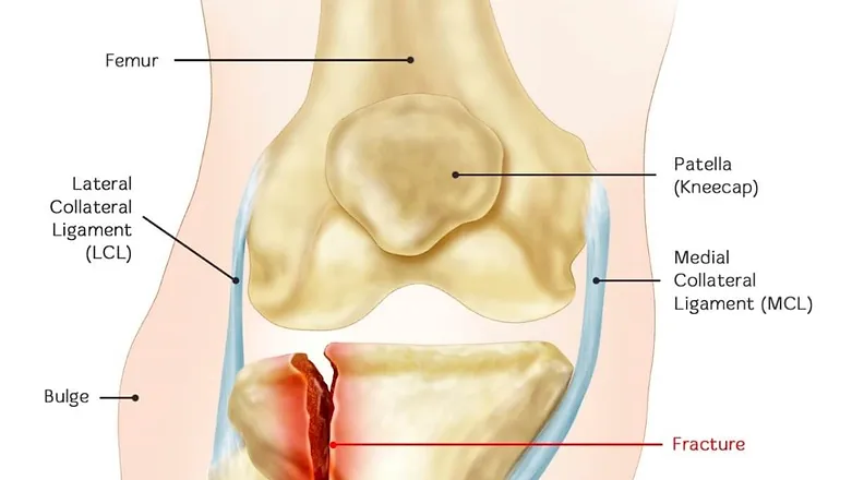
A fracture, or break, in the shinbone just below the knee is called a proximal tibia fracture. The proximal tibia is the upper portion of the bone where it widens to help form the knee joint.
In addition to the broken bone, soft tissues (skin, muscle, nerves, blood vessels, and ligaments) may be injured at the time of the fracture. Both the broken bone and any soft-tissue injuries must be treated together. In many cases, surgery is required to restore strength, motion, and stability to the leg, and reduce the risk for arthritis.
There are several types of proximal tibia fractures. The bone can break straight across (transverse fracture) or into many pieces (comminuted fracture).
Sometimes these fractures extend into the knee joint and separate the surface of the bone into a few (or many) parts. These types of fractures are called intra-articular or tibial plateau fractures.
The top surface of the tibia (the tibial plateau) is made of cancellous bone, which has a “honeycombed” appearance and is softer than the thicker bone lower in the tibia. Fractures that involve the tibial plateau occur when a force drives the lower end of the thighbone (femur) into the soft bone of the tibial plateau, similar to a die punch. The impact often causes the cancellous bone to compress and remain sunken, as if it were a piece of Styrofoam that has been stepped on.
This damage to the surface of the bone may result in improper limb alignment and, over time, may contribute to arthritis, instability, and loss of motion.
Proximal tibia fractures can be closed — meaning the skin is intact — or open. An open fracture is when a bone breaks in such a way that bone fragments stick out through the skin or a wound penetrates down to the broken bone. Open fractures often involve much more damage to the surrounding muscles, tendons, and ligaments. They have a higher risk for problems like infection, and take a longer time to heal.
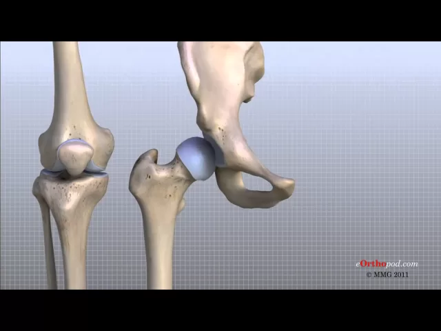
KNEE ANATOMY
DISEASE EXPLAINED

SYMPTOMS
Pain that is worse when weight is placed on the affected leg
Swelling around the knee and limited bending of the joint
Deformity — The knee may look “out of place”
Pale, cool foot — A pale appearance or cool feeling to the foot may suggest that the blood supply is in some way impaired.
Numbness around the foot — Numbness, or “pins and needles,” around the foot raises concern about nerve injury or excessive swelling within the leg.
If you have these symptoms after an injury, go to the nearest hospital emergency room for an evaluation.
CAUSES
Patellar fractures are most often caused by:
Falling directly onto the knee
Receiving a sharp blow to the knee, such as might occur during a head-on vehicle collision if your kneecap is driven into the dashboard
The patella can also be fractured indirectly. For example, a sudden contraction of the quadriceps muscle in the knee can pull apart the patella.
TREATMENT
TREATMENT OPTIONS
A proximal tibia fracture can be treated nonsurgically or surgically. There are benefits and risks associated with both forms of treatment.
Whether to have surgery is a combined decision made by the patient, the family, and the doctor. The preferred treatment is accordingly based on the type of injury and the general needs of the patient.
When planning treatment, your doctor will consider several things, including your expectations, lifestyle, and medical condition.
In an active individual, restoring the joint through surgery is often appropriate because this will maximize the joint’s stability and motion, and minimize the risk of arthritis.
In other individuals, however, surgery may be of limited benefit. Medical concerns or pre-existing limb problems might make it unlikely that the individual will benefit from surgery. In such cases, surgical treatment may only expose these individuals to its risks (anesthesia and infection, for example).
EMERGENCY CARE
OPEN FRACTURES.
If the skin is broken and there is an open wound, the underlying fracture may be exposed to bacteria that might cause infection. Early surgical treatment will cleanse the fracture surfaces and soft tissues to lessen the risk of infection.
EXTERNAL FIXATION.
If the soft tissues (skin and muscle) around your fracture are badly damaged, or if it will take time before you can tolerate a longer surgery because of health reasons, your doctor may apply a temporary external fixator. In this type of operation, metal pins or screws are placed into the middle of the femur (thighbone) and tibia (shinbone). The pins and screws are attached to a bar outside the skin. This device holds the bones in the proper position until you are ready for surgery.
COMPARTMENT SYNDROME.
In a small number of injuries, soft-tissue swelling in the calf may be so severe that it threatens blood supply to the muscles and nerves in the leg and foot. This is called compartment syndrome and may require emergency surgery. During the procedure, called a fasciotomy, vertical incisions are made to release the skin and muscle coverings. These incisions are often left open and then stitched closed days or weeks later as the soft tissues recover and swelling resolves. In some cases, a skin graft is required to help cover the incision and promote healing.
NONSURGICAL TREATMENT
Nonsurgical treatment may include casting and bracing, in addition to restrictions on motion and weight bearing. Your doctor will most likely schedule additional x-rays during your recovery to monitor whether the bones are healing well while in the cast. Knee motion and weight-bearing activities begin as the injury and method of treatment allow.
SURGICAL TREATMENT
There are a few different methods that a surgeon may use to obtain alignment of the broken bone fragments and keep them in place while they heal.
INTERNAL FIXATION.
During this type of procedure, the bone fragments are first repositioned (reduced) into their normal position. They are held together with special devices, such as an intramedullary rod or plates and screws.
In cases in which the upper one fourth of the tibia is broken, but the joint is not injured, a rod or plate may be used to stabilize the fracture. A rod is placed in the hollow medullary cavity in the center of the bone. A plate is placed on the outside surface of the bone.
Plates and screws are commonly used for fractures that enter the joint. If the fracture enters the joint and pushes the bone down, lifting the bone fragments may be required to restore joint function. Lifting these fragments, however, creates a hole in the cancellous bone of the region. This hole must be filled with material to keep the bone from collapsing. This material can be a bone graft from the patient or from a bone bank. Synthetic or naturally occurring products which stimulate bone healing can also be used.
EXTERNAL FIXATORS.
In some cases, the condition of the soft tissue is so poor that the use of a plate or rod might threaten it further. An external fixator (described under Emergency Care above) may be considered as final treatment. The external fixator is removed when the injury has healed.
DISCLAIMER
- All of the opinions expressed within the educational information delivered within the provided links are those of their authors and not necessarily those of either your treating doctor or CRO.
- This site is for educational purposes only!!
- Copyright Disclaimer under Section 107 of the copyright act 1976, allowance is made for fair use for purposes such as criticism, comment, news reporting, scholarship, and research. Fair use is a use permitted by copyright statutes that might otherwise be infringing. Non-profit, educational, or personal use tips the balance in favor of fair use.